
Analysis of Human Papillomavirus (HPV) Detection by p16 Immunoreactivity in External Hemorrhoids
Análise da presença de HPV por imunorreatividade para p16 em hemorroidas externas
Júlia Yumi Fujiki1, Sofia Alessandra Kotsifas1, Laura Mendes Lopes1, Jennifer Thais de Barros Ferro1, Ana Cristina Lira Sobral1
1 Faculdade Evangélica Mackenzie do Paraná. Curitiba, PR, Brazil.
Received on 10/07/2024
Approved on 01/03/2025
DOI: 10.21877/2448-3877.202500201.en
INTRODUCTION
Human papillomavirus (HPV) is a double-stranded DNA virus belonging to the Papillomaviridae family(1) that exhibits tropism for epithelial tissues(2) by exploiting cellular renewal to enter the epithelium and infect basal layer cells.(3) HPV is the most common sexually transmitted infection (STI) worldwide.(4)
HPV types are classified as low-risk, responsible for anogenital and cutaneous warts, or high-risk, which can lead to the development of anogenital and oropharyngeal cancers.(4)
These high-risk types express the oncoproteins E6 and E7, which promote cell proliferation and inactivate cell-cycle checkpoints by interfering with the regulatory proteins p53 and pRb.(3,5) Thus, HPV proteins can drive epithelial tissue toward neoplastic phenotypes,(6) which may also occur in hemorrhoidal tissue, a site that favors HPV tropism. The virus is frequently detected incidentally during pathological analysis of tissue removed during hemorrhoidectomy.
The gold-standard treatment for external hemorrhoids (EH) is hemorrhoidectomy, and current guidelines recommend a three-year follow-up after surgery to assess the potential for hemorrhoidal recurrence. However, HPV may be present in the tissue, representing a risk factor for the development of anorectal cancer and potentially remaining latent in the residual tissue for up to five years. Therefore, histopathological evaluation of hemorrhoidectomy specimens is warranted, allowing extended follow-up for HPV-positive patients to cover the virus’s longer latency period in hemorrhoidal tissue.
OBJECTIVE
To assess p16 marker positivity in external hemorrhoidal tissue and analyze its distribution by age group and sex.
MATERIALS AND METHODS
This observational study reviewed histopathology reports from 44 patients who underwent hemorrhoidectomy and showed cellular alterations consistent with koilocytosis, identified under optical microscopy (OM).
After selection, the areas showing these cellular alterations were demarcated and localized using a mirror system, in which the marked slide was used to identify the corresponding region in the donor block. A perforation was then made in the donor block at the previously marked area to extract a tissue core, which was subsequently implanted into the recipient block (Figure 1A). Each sampled fragment removed from the donor block was placed in the recipient block following a “Cartesian plane” mapping scheme (Figure 1B). In this manner, multi-sample tissue blocks (TMAs) were constructed (Figure 1C), which were then used to prepare multi-sample slides containing representative lesional areas (Figure 1D).
Histological slides obtained from the handcrafted TMA blocks were subjected to the immunohistochemistry process for the p16 marker in two phases: application of the standardized immunohistochemical technique to the study material, followed by microscopic reading of the positive areas on the slides. Two liver slides immunostained with the p16 antibody served as controls and reference standards for defining the mean area of marker expression across the series. Subsequently, the microscopic readings were tabulated in an Excel spreadsheet and submitted for statistical analysis.
Continuous variables were expressed as mean ± standard deviation and compared using Student’s t-test or the Mann-Whitney test. Categorical variables were expressed as percentages and compared using the chi-square or Fisher’s exact test, as appropriate. p-values < 0.05 were considered statistically significant.
Patients were classified as p16-positive or p16-negative following immunohistochemical analysis, and these findings were correlated with prevalence by sex and age group, divided into three categories: 18–25, 26–35, and ≥ 36 years.
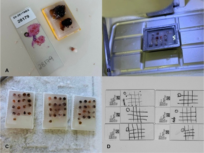
Figure 1
Preparation of tissue sample slides
RESULTS
Hemorrhoidal mucosal samples were collected from 44 patients presenting koilocytotic cellular alterations indicative of HPV infection, of whom 26 were female and 18 were male. Two patients were aged 18–25 years (4.55%), five were aged 26–35 years (11.36%), and 37 were aged ≥ 36 years (84.09%). The mean age was 48.34 years. Of the 44 patients, 21 showed hemorrhoidal tissue staining positive for p16 immunohistochemistry, indicating potential oncogenic progression (47.73%) (Figure 2).
The mean age of p16-positive cases was 51.62 years, whereas negative cases had a mean age of 45.35 years. There was no statistically significant difference between the groups regarding age (p = 0.13) (Table 1).
Among the 26 female patients, nearly 54% tested positive, whereas approximately 39% of male patients tested positive. However, this difference was not statistically significant (p = 0.33).
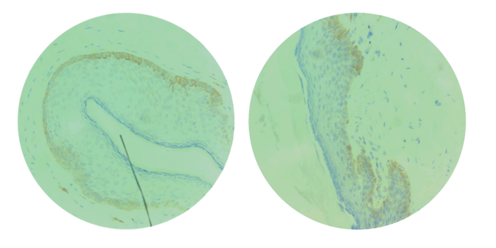
Figure 2
Analysis of p16 positivity.
*Hemorrhoidal tissue sample for p16 (brown area ×400)
Table 1
Relationship between age and p16 positivity.
| Age | |||||
| Positivity | Mean | n | SD | Minimum | Maximum |
| Yes | 51.62 | 21 | 14.86 | 25 | 96 |
| No | 45.35 | 23 | 11.44 | 20 | 68 |
| Total | 48.34 | 44 | 13.41 | 20 | 96 |
SD: standard deviation. Source: Authors, 2023.
DISCUSSION
HPV infection is associated with squamous cell carcinoma and intraepithelial neoplasia of the anal canal, perianal region, vulva, vagina, and penis.(7) Hemorrhoidal tissue, due to its squamous epithelial composition, may have its basal cells targeted by HPV infection, which may act as a predisposing factor for dysplasia and neoplastic transformation.(8,9) Approximately 48% of patients with external hemorrhoids who underwent hemorrhoidectomy and presented cellular alterations compatible with koilocytosis showed immunohistochemical staining for the p16 marker, indicating potential oncogenic risk associated with HPV presence in hemorrhoidal tissue.
According to the meta-analysis by Pelizzer et al. (2016),(10) the relationship between colorectal cancer and HPV is strong: HPV was detected in 51.5% of the colorectal cancers analyzed, with most cases associated with HPV types 16 and 18, and a similar incidence observed in both sexes. Confirmation of the studied neoplasms was more frequently established at advanced stages, likely due to late diagnosis, which may be explained by failures in the screening methods for this condition.(10)
Through p16 immunohistochemistry, this study found that 47.73% (21/44) of patients who underwent hemorrhoidectomy showed p16 positivity. This finding differs somewhat from that of Jian-xiang Geng et al. (2011),(11) who used quantitative fluorescent PCR to detect HPV and reported that 69.6% of their external hemorrhoid samples were HPV-positive. This discrepancy may be explained by the sensitivity and specificity of the different techniques used, as quantitative PCR, used by Jian-xiang Geng et al. (2011) (11), is highly sensitive and can detect the virus even when present in small quantities, whereas p16 immunohistochemistry, used in the present study, is particularly effective at detecting HPV-induced cellular alterations that usually progress to lesions with high oncogenic potential.(12,13)
The choice of the p16 marker for immunohistochemistry in this study was based on its high negative predictive value. Thus, the absence of elevated p16 expression in tissue samples is highly indicative of a lack of risk for HPV-related dysplasia, making p16 particularly important for screening and identifying lesions.(14) It plays a crucial role in clinical practice by helping determine which patients can be safely monitored over time without the need for invasive procedures, given that hemorrhoidectomy is a less aggressive procedure than resection of anorectal tissue. Furthermore, the presence of HPV infection in hemorrhoidal tissue increases the likelihood of similar alterations in other susceptible tissues.(15)
The study by Lu DW et al. (2003),(16) conducted on anorectal squamous cell carcinoma, found 100% cytoplasmic p16 positivity in their cases, whereas the adjacent non-neoplastic squamous and mucosal epithelium showed no evident nuclear staining. Therefore, in this study, negative samples (52.27%) likely exclude HPV as a contributing risk factor for the development of anorectal carcinoma.
For an analysis based on age groups, patients were divided into three categories: 18–25 years, 26–36 years, and >36 years. It was observed that just over 84% of patients with external hemorrhoidal disease (EHD) were aged 36 years or older, with a mean age of 48.34 years (ranging from 20 to 96 years), corroborating the findings of Cristea and Lewis (2023),(17) who reported that the most prevalent age range for EHD is 45–65 years. However, although advanced age is a risk factor for the development of external hemorrhoids (EH), this study demonstrated no significant relationship between age and p16 positivity (p = 0.25). Therefore, it is not possible to associate the coexistence of HPV in the tissue with the patient’s age, implying that screening for HPV infection in hemorrhoidal tissue should be offered to all patients undergoing hemorrhoidectomy for EHD, regardless of age.
Similarly, regarding the distribution of EHD by sex, it was observed that of the 44 patients included in the study, 59.09% (26) were female, a percentage similar to that found in a previous Brazilian study,(9) which reported an incidence of 53.9% in women, demonstrating that sex is not a risk factor for the development of EHD. Furthermore, when analyzing p16 expression in the excised hemorrhoidal tissue, nearly 54% of female patients tested positive, compared with approximately 39% of male patients. Although positivity was more prevalent among females in this study, the difference between sexes was not statistically significant (p = 0.33), reinforcing that the neoplastic risk associated with the coexistence of HPV in the tissue cannot be attributed to patient sex. In agreement with these findings, other studies on HPV have also shown no difference in infection incidence between sexes.(10) Therefore, patient sex should not be considered a determining factor when planning screening for potential HPV infection in hemorrhoidal tissue.
Study Limitations
As this was a retrospective observational study, patient risk behaviors related to HPV infection could not be assessed; therefore, it was not possible to determine whether these patients might have had higher rates of HPV-related lesions in hemorrhoidal tissue. Nevertheless, even if this assumption were true, there is no evidence suggesting that these patients would require a different post-hemorrhoidectomy follow-up protocol than that recommended for other patients.
Furthermore, due to the retrospective nature of the study, it was not possible to assess the incidence of anorectal carcinoma in patients who were not followed clinically for the maximum HPV latency period in hemorrhoidal tissue, that is, five years after the procedure.
This study did not analyze slides containing samples of internal rectal tissue. Therefore, it cannot be stated that the presence of koilocytotic changes in external hemorrhoidal tissue necessarily indicates similar alterations in internal tissue, which could serve as a risk factor for the development of precursor lesions for anorectal cancer. In addition, the high negative predictive value of the p16 marker in HPV-infected tissue, used to analyze the samples, reduces the likelihood of undetected lesions. Nevertheless, there is no reason to suggest that patients with p16 positivity would benefit from a different post-hemorrhoidectomy follow-up than that recommended for other patients, as HPV screening plays an important role in the early detection of preneoplastic lesions and, consequently, in cancer prevention.
Clinical Implications
Given that the standard management of external hemorrhoids involves hemorrhoidectomy followed by a three-year postoperative clinical follow-up, and that HPV can remain latent in tissue for up to five years, a careful evaluation of hemorrhoidal tissue is warranted. In cases where microscopic examination reveals cells with eccentric nuclei and a clear perinuclear halo, features indicative of HPV infection, post-hemorrhoidectomy follow-up should be extended to investigate the risk of neoplastic development associated with HPV in the internal anorectal tissue, thus accommodating the virus’s potential latency period.
This discrepancy between the HPV latency period in hemorrhoidal tissue and the duration of standard postoperative follow-up justifies adopting a more proactive investigative protocol focused on early diagnosis, treatment, and prevention of these lesions, thereby reducing the likelihood of neoplasm development or enabling timely clinical management.
CONCLUSION
The data obtained in this study demonstrate that among patients who underwent hemorrhoidectomy and presented microscopic findings resembling koilocytes, indicative of HPV, approximately half showed p16 positivity, with neither age nor sex emerging as risk factors.
Accordingly, it is emphasized that patients undergoing hemorrhoidectomy who exhibit such microscopic findings may benefit from p16 testing and extended follow-up to detect precancerous lesions.
REFERENCES
- Mateos-Lindemann ML, Pérez-Castro S, Rodríguez-Iglesias M, Pérez-Gracia MT. Diagnóstico microbiológico de la infecção por vírus do papiloma humano. Enferm Infecc Microbiol Clin. 2017;35(9):593-602.
- Magalhães GM, Vieira EC, Garcia LC, de Lourdes Ribeiro De Carvalho-Leite M, Guedes ACM, Araújo MG. Update on human papilloma virus – part I: epidemiology, pathogenesis, and clinical spectrum. An Bras Dermatol. 2021;96(1):1-6.
- McBride AA. Oncogenic human papillomaviruses. Philos Trans R Soc Lond B Biol Sci. 2017;372(1732):1-9.
- Kombe AJK, Li B, Zahid A, Mengist HM, Bounda G, Zhou Y, et al. Epidemiology and burden of human papillomavirus and related diseases, molecular pathogenesis, and vaccine evaluation. Front Public Health. 2020;8:552028.
- Monsonego J. Cancer anal et papillomavirus humains: une pathologie en miroir de celle du cancer du col utérin. Gynécologie Obstétrique & Fertilité. 2010;38(4):250-254.
- Kranjec C, Doorbar J. Human papillomavirus infection and induction of neoplasia: a matter of fitness. Curr Opin Virol. 2016;20:129-136.
- Mahajan A. Practical issues in the application of p16 immunohistochemistry in diagnostic pathology. Hum Pathol. 2016;51:64-74. doi:10.1016/j.humpath.2015.12.021.
- Petca A, Borislavschi A, Zvanca ME, Petca R, Sandru F, Dumitrascu MC. Non-sexual HPV transmission and role of vaccination for a better future (Review). Exp Ther Med. 2020;20(6):1-5.
- Da Cruz GM, Ferreira RMRS, Neve PM. Doença hemorroidária: aspectos epidemiológicos e diagnósticos de 9.289 pacientes portadores de doença hemorroidária. Rev Bras Colo-Proctol. 2006;26(1):6-23.
- Pelizzer T, Dias CP, Poeta J, Torriani T, Roncada C. Prevalência de câncer colorretal associado ao papilomavírus humano: uma revisão sistemática com metanálise. Rev Bras Epidemiol. 2016;19(4):791-802.
- Geng JX, Fan ZM, Ding YJ, Chen JN, Zhang SM, Zhou W, Huang SL, Han DM. [Analysis on human papillomavirus 16 and 18 types infection among 805 patients with common anorectal lesions]. Zhonghua Wei Chang Wai Ke Za Zhi. 2011;14(12):958-60.
- Castello F, Vidal B, Do M, Soares D, Nascimento B, Tereza C, et al. Análise crítica dos métodos moleculares para detecção do papilomavírus humano: revisão da literatura. Rev Bras Patol Med Lab. 2012;40(5).
- Yonamine P, Junqueira M, Rodrigues J, Pereira S, Pandossio T, Rodrigues D, et al. Associação entre a expressão de p16 e neoplasia intra-epitelial cervical. Arq Cienc Saude. 2009;16(4):161-6.
- Salim RC, Lima TM, Heinke T, Prigenzi KCK, Speck NMG, Focchi GRA. Cervical squamous intraepithelial lesions grades 1 and 2: value of an alternative morphological classification and of p16 immunohistochemistry in the prediction of clinical outcome. J Bras Patol Med Lab. 2017;53(5):313-9.
- Gonçalves JES, et al. The role of p16 as putative biomarker for cervical neoplasia: A controversial issue? MedExpress. 2017;4(6).
- Lu DW, El-Mofty SK, Wang HL. Expression of p16, Rb, and p53 proteins in squamous cell carcinomas of the anorectal region harboring human papillomavirus DNA. Mod Pathol. 2003;16(7):692-9. doi:10.1097/01.MP.0000077417.08371.CE.
- Cristea C, Lewis CR. Hemorrhoidectomy. In: StatPearls [Internet]. Treasure Island (FL): StatPearls Publishing; 2023.
Correspondence
Júlia Yumi Fujiki
E-mail: [email protected]