
Cryoglobulin crystals in peripheral blood
Cristais de crioglobulina em sangue periférico
Samuel Ricardo Comar1, Marcio Augusto Batista1, Salomão Cury Riechi1
1 Universidade Federal do Paraná, Laboratório de Hematologia do Complexo Hospital de Clínicas. Curitiba, PR, Brazil.
Received on 05/17/2025
Approved on 07/02/2025
DOI: 10.21877/2448-3877.202500244.en
Cryoglobulin crystals in peripheral blood
Cryoglobulinemia is a rare condition characterized by the presence of abnormal immunoglobulins in plasma that precipitate at temperatures below 37 °C and redissolve upon rewarming. Although classically associated with clinical manifestations such as purpura, arthralgia, vasculitis, glomerulonephritis, and peripheral neuropathy, it may also be related to chronic lymphoproliferative disorders. The occurrence of visible crystals in peripheral blood smears is extremely uncommon and rarely reported in the literature.
In the images, we describe a unique case of cryoglobulinemia evidenced by amorphous, gray-blue, weakly basophilic, needle-like structures observed in a peripheral blood smear stained with May-Grünwald-Giemsa and examined under light microscopy. The morphology of these extracellular crystals, in conjunction with the clinical context and compatible laboratory findings, such as elevated serum proteins, the presence of rheumatoid factor, and a positive cryoglobulin test, allows for an accurate correlation with the underlying pathology. These findings may also serve as an important diagnostic clue in patients presenting with nonspecific clinical manifestations.
Cryoglobulins can interfere with automated hematologic analyses, resulting in pseudoleukocytosis, pseudothrombocytosis, and abnormal histograms and scatter plots. Therefore, recognizing these hematologic abnormalities and cryoglobulin-induced artifacts is essential, as they may constitute the first indication leading to the timely diagnosis of cryoglobulinemia.
This finding underscores the role of the hematology laboratory and the careful morphological evaluation of blood smears as fundamental tools in the investigation of certain immunoproliferative and autoimmune diseases, even in the era of automated multiparametric hematology analyzers.
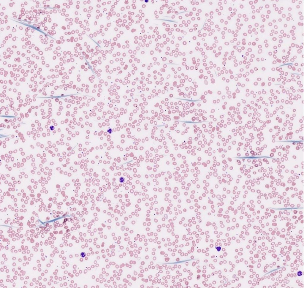
Figure 1
Cryoglobulin crystals in peripheral blood.
Peripheral blood smear stained with May-Grünwald-Giemsa showing multiple elongated structures consistent with cryoglobulin crystals, scattered among normocytic red blood cells and preserved leukocytes.
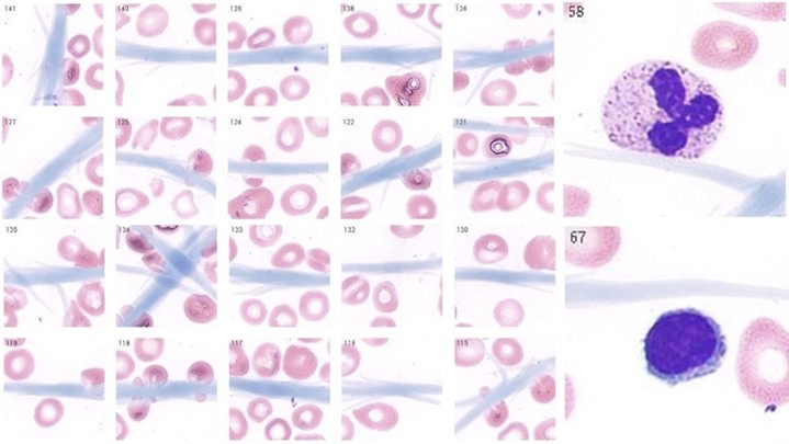
Figure 2
Cryoglobulin crystals at high magnification: atypical extracellular structures in peripheral blood smear.
High-power view of a peripheral blood smear stained with May-Grünwald-Giemsa showing cryoglobulin crystals as bluish, elongated, or rosette-like extracellular structures. These rare morphological findings are highly suggestive of cryoglobulinemia and may serve as a key diagnostic clue, particularly in patients with nonspecific clinical presentations.
REFERENCES
- Dave RG, Padiyar S, Mathew J, Nair SC. Unusual morphological and automated hematology analyzer features in 3 cases of B-cell malignancy-associated type I cryoglobulinemic Vasculitis. Indian J Hematol Blood Transfus. 2021;37(4):658-663. doi:10.1007/s12288-021-01398-w
- Wang F, Wang G, Wang X. Cryoglobulin crystals deposited in a peripheral blood film and phagocytosed by neutrophils. Br J Haematol. 2021;195(4):484. doi:10.1111/bjh.17672
- Mohd Zaini WS, Hamour S, Swamy R, Kumar P. Type 1 cryoglobulinaemia with precipitated crystals. Br J Haematol. 2013;161(3):301. doi:10.1111/bjh.12243
Correspondence
Samuel Ricardo Comar
E-mail: [email protected]