
Spheroplasts in Urinary Sediment: Bacterial Forms Associated with Exposure to Beta-Lactams
Esferoplastos no Sedimento Urinário: Formas Bacterianas Associadas à Exposição a β-Lactâmicos
Charben Abdala de Paula Silva1, Ana Paula Giolo Franz2, Mauren Isfer Anghebem3
1 Biomédico; Especialização em Hematologia Laboratorial e Clínica; Responsável Técnico no Laboratório do Hospital e Maternidade Dona Lisette. Taió, SC, Brazil.
2 Biomédica, Doutora em Ciências, Responsável Técnica do Laboratório de Análises Clínicas do Hospital de Clínicas Mais Saúde. Passo Fundo, RS, Brazil. Membro do Grupo Técnico-Científico da Sociedade Brasileira de Análises Clínicas – SBAC.
3 Farmacêutica-Bioquímica, Doutorado em Ciências Farmacêuticas/Análises Clínicas; Título de Especialista em Análises Clínicas – SBAC; Professora do Departamento de Análises Clínicas da Universidade Federal do Paraná e da Escola de Medicina e Ciências da Vida da Pontifícia Universidade Católica do Paraná. Curitiba, PR, Brazil.
Received on 07/31/2026
Approved on 07/31/2026
DOI: 10.21877/2448-3877.202600330.en
Spheroplasts are bacterial forms with partially deficient cell walls that can be observed in urinary sediment, usually associated with exposure to subinhibitory concentrations of β-lactam antibiotics. Microscopically, they appear as spherical structures of varying sizes, often accompanied by filamentous extensions or irregular protrusions, which may aid in their differentiation from erythrocytes and fungal hyphae. While hyphae exhibit typical septation and branching, spheroplasts display non-septate filaments in addition to marked size variability.
Bacterial morphological integrity depends on the peptidoglycan of the cell wall, the synthesis of which is mediated by penicillin-binding proteins (PBPs). β-lactams act by binding to these proteins and inhibiting cell wall synthesis. At adequate concentrations, bacterial lysis occurs; however, at subinhibitory levels, there may be partial loss of the cell wall, resulting in filamentation or transformation into spherical forms called spheroplasts. PBP 2 is essential for the synthesis of the lateral cell wall. When inhibited, the cell loses its rod-shaped form and becomes spherical, resulting in spherulation and, eventually, osmotic lysis. PBP 3, on the other hand, is necessary for murein synthesis and cell division. Its inhibition prevents the cell from dividing but allows it to continue growing, forming long, multinucleated filaments.
In the urinary tract, these forms can remain viable and relatively stable, particularly under favorable physicochemical conditions. Studies have demonstrated greater stability of Escherichia coli and Proteus vulgaris spheroplasts in media with a pH between 5.0 and 5.5. Furthermore, acidic and relatively hyperosmolar urine favors their persistence and reduces the osmotic lysis of these fragile forms. The ability to revert to the bacillary form after antibiotic discontinuation suggests a potential role in episodes of bacterial persistence and urinary reinfection.
From a laboratory perspective, spheroplasts are of high diagnostic significance, especially given the growing use of automated urine sediment analyzers based on image-based morphological recognition. These structures may be misclassified as red blood cells, yeast, or cellular debris, mainly due to their similarity in size and shape. A discrepancy between a high automated red blood cell count and a negative hemoglobin test on the test strip should prompt manual microscopic review. In this context, phase-contrast microscopy can significantly contribute to the morphological characterization of these structures.
The correct identification of spheroplasts in urinary sediment may represent an indirect marker of exposure to subinhibitory concentrations of β–lactams, treatment failure, or bacterial resistance, including infections caused by β-lactamase-producing Enterobacteriaceae, such as ESBL or KPC producing Klebsiella pneumoniae. Thus, their recognition by the clinical laboratory has the potential to impact the interpretation of urine cultures, the assessment of therapeutic efficacy, and communication with the healthcare team.
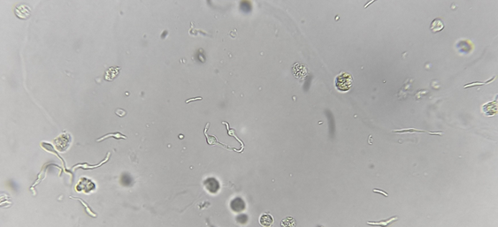
Figure 1
Urinary sediment under bright-field microscopy showing spherical and filamentous structures of varying sizes consistent with bacterial spheroplasts.
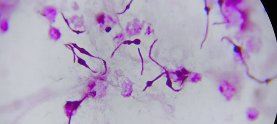
Figure 2
The same urinary sediment, viewed under a microscope after Gram staining.
Bibliography
Falbo R, Fogazzi GB, Sala MR, Garigali G, Sulejmani A, Brambilla P, et al. Spheroplasts, poorly known but clinically relevant particles of urinary sediment. Clin Chim Acta. 2021;515:13-15.
Nikler A, Radišić Biljak V, Čičak H, Marić N, Bejuk D, Poloni JAT, et al. Escherichia coli spheroplasts in a Croatian patient misclassified by two urine sediment analysers as erythrocytes: case report. Biochem Med (Zagreb). 2019;29(3):030801.
Poloni JAT, Meinerz G, Monteiro AA, Keitel E, Rotta LN. Klebsiella pneumoniae ESBL forming spheroplasts in the fresh and unstained urine sediment. J Bras Nefrol. 2016;38(2):269-270.
Correspondence
Mauren Isfer Anghebem
E-mail: [email protected]