
Use of MicroRNAs as an intervention in the early diagnosis of breast cancer: a systematic review
Emprego de MicroRNAs como intervenção no diagnóstico precoce do câncer de mama: uma revisão sistemática
Bruna Kühnel1, Cristian Roncada2, Marco Antônio Ghiotto3
1,2 Centro Universitário da Serra Gaúcha, Biomedicina. Caxias do Sul, RS, Brazil.
3 Universidade FEEVALE, Biomedicina. Novo Hamburgo, RS, Brazil.
Received on 02/18/2025
Approved on 07/02/2025
DOI: 10.21877/2448-3877.202500223.en
INTRODUCTION
Breast cancer (BC) is the most common malignant neoplasm affecting women in Brazil and worldwide. According to data from the Brazilian National Cancer Institute (INCA), it is the leading cause of cancer-related death among women, second only to non-melanoma skin tumors in terms of incidence. Updated annual estimates highlight the evolving patterns of incidence and mortality across regions, characterizing BC as a serious public health concern. In Brazil, the Southern Region reports the highest rates of the disease, with the state of Santa Catarina registering the highest age-adjusted incidence of new cases. Santa Catarina, followed by Rio Grande do Sul and Paraná, also reports elevated BC mortality rates. This high incidence is attributed to factors such as increased awareness of the disease and improved access to diagnosis and appropriate treatment.(1,2)
Although BC is rare before the age of 30, the risk increases progressively with age. However, studies indicate a growing prevalence of the disease among younger women. The main symptoms include breast or axillary lumps, breast pain, skin or nipple retraction, swelling, and weight loss.(3) Early detection is essential for a better prognosis, with mammography being the gold-standard screening method, recommended for women within specific age groups and performed at regular intervals.(4)
Delayed diagnosis of breast cancer can lead to serious consequences, significantly increasing the risk of mortality. Survival rates vary according to several factors, including nutritional status, immune function, patient age, and the tumor’s metastatic potential. Young women and pregnant patients generally present lower survival rates. BC treatment includes surgery, radiotherapy, hormone therapy, and chemotherapy, with early identification of malignancy being critical to therapeutic success.(5,6)
In this context, microRNAs (miRNAs) have emerged as key regulators of biological processes such as cell proliferation, apoptosis, and differentiation, directly influencing the development and progression of BC. miRNA-encoding genes are often located in unstable chromosomal regions and can be detected in plasma or serum, enabling differentiation between healthy individuals and those with cancer. This feature makes miRNAs promising biomarkers for early disease detection.(7,8)
Given these considerations, the aim of the present study is to contribute to the improvement of breast cancer screening and diagnostic programs by providing a more precise and effective approach to identifying the disease in its early stages, thereby expanding treatment options and survival rates, and offering a new perspective in the fight against breast cancer.
METHODS
This systematic literature review was registered under the registration number CRD42024566138 in the International Prospective Register of Systematic Reviews (PROSPERO)(9) and followed the guidelines of the Preferred Reporting Items for Systematic Reviews and Meta-Analyses (PRISMA)(10).
The search strategy employed the combination of descriptors “MicroRNAs OR miRNAs AND Breast Cancer Diagnosis”, applied to MEDLINE (PubMed), SciELO (BIREME), and ScienceDirect (Elsevier). A publication date filter was applied to include studies published from 2010 onward. For validation purposes, the search logic used on PubMed was: (MicroRNAs[Title/Abstract] OR MicroRNAs[MeshTerms] OR miRNAs[Title/Abstract] OR miRNAs[MeshTerms]) AND (“Breast Cancer”[Title/Abstract] OR “Breast Cancer”[MeshTerms]) AND (Diagnosis[Title/Abstract] OR Diagnosis[MeshTerms]).
After exporting the selected studies from the databases, the StArt software (State of the Art through Systematic Review)(11) was used to generate the study flowchart, comprising four phases: a) Identification – recruitment of studies; b) Screening – removal of duplicates and exclusion based on title and abstract review; c) Eligibility – exclusion based on full-text review; d) Inclusion – studies meeting the predefined inclusion criteria.
Article selection was conducted in three independent phases by two researchers and reviewed by a third collaborator. The inclusion criteria were as follows: articles selected by both researchers during the initial screening were directly included in the review; additionally, studies identified through reference lists of already included articles during the eligibility phase were also considered. Conversely, exclusion criteria involved articles not selected by either researcher, which were automatically discarded. In cases where an article was selected by only one researcher, it was submitted for evaluation by the third collaborator, who made the final decision regarding its inclusion or exclusion.
The search was conducted between August and September 2024. For data extraction and identification of eligible studies, standardized data-extraction tables were used to record information such as the first author, year of publication, clinical trial period and data collection, country of origin, number of participants, and study design.
The inclusion criteria for the systematic review encompassed randomized clinical trials, non-randomized clinical trials, case reports, comparative studies, and case-control studies that investigated the use of microRNAs for the early diagnosis of breast cancer.
RESULTS
A total of 210 articles were initially selected: 22 from the PubMed (MEDLINE) database, 180 from ScienceDirect (Elsevier), 2 from SciELO (BIREME), and 6 identified through grey literature. The selection of these additional articles followed the same eligibility criteria applied to the other studies and were identified through reference searches of included articles and expert recommendations to fill gaps identified in the preliminary analysis.
Among these, 3 articles were excluded due to duplication across databases. Thus, from the remaining 207 articles, 180 were excluded based on title and abstract screening, leaving 21 articles for full-text review. All 21 were deemed eligible for inclusion in the systematic review, as shown in Figure 1.
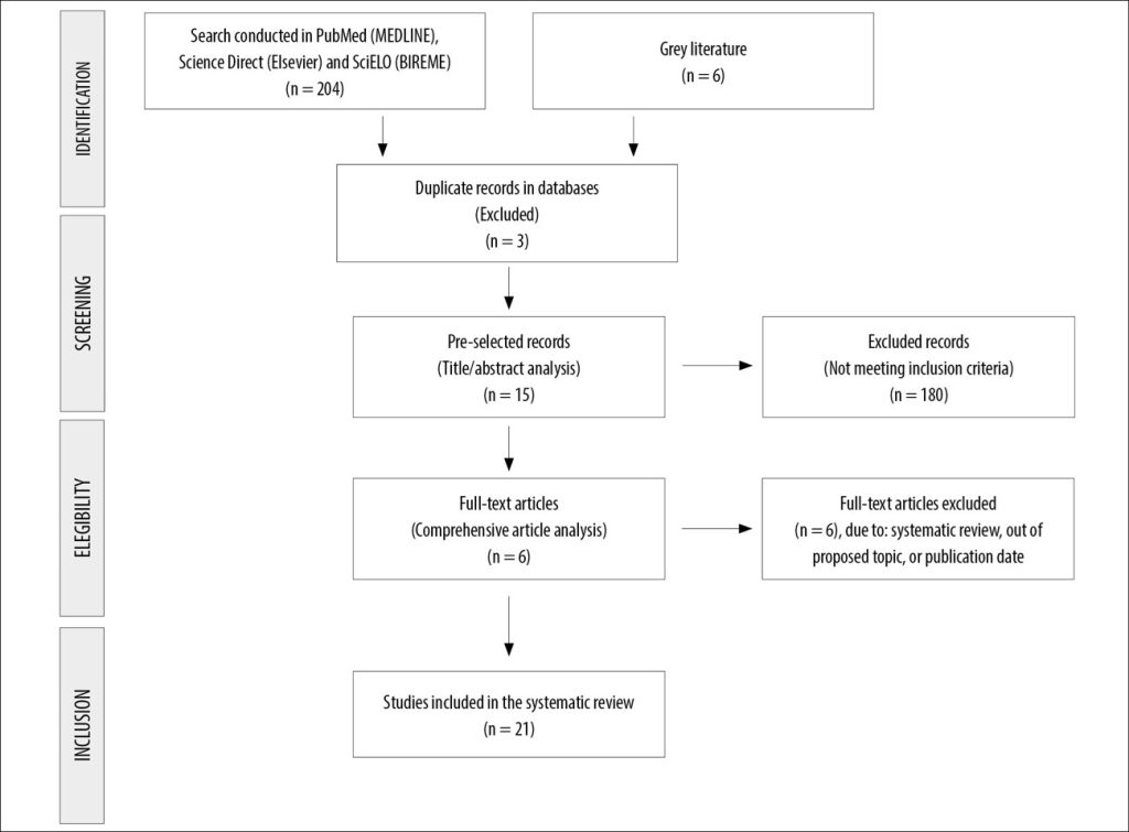
Figure 1
Flowchart of articles eligible for the systematic review.
Source: Adapted from PRISMA.
Among the 21 studies included in the systematic review, the country distribution revealed that China accounted for the highest number of publications (n = 6; 28.6%),(11,14,15,24,25,30) indicating strong scientific interest in the application of microRNAs (miRNAs) for early breast cancer diagnosis. Egypt (n = 4)(17,21,23,26) and Germany (n = 3)(12,13,19) also stood out, reflecting significant contributions from both developing and European countries.
Regarding study design, case-control studies predominated (n = 9),(12-14,16,20,21,23,26,29) followed by observational studies (n = 7),(18,19,24,25,27,28,30) retrospective studies (n = 2),(17,31) one clinical study, (23) and one cohort study.(11) This distribution shows a clear preference for designs aimed at identifying associations between miRNA expression and breast cancer occurrence, with case-control studies being particularly suitable for diagnostic evaluation.
The total number of participants varied considerably across studies. The study by Enders et al.(15) included 525 participants, contributing to greater statistical robustness. Altogether, the 21 studies comprised 1,871 participants. The mean age reported in studies that included this variable was approximately 53.3 years, consistent with the age range most affected by breast cancer.
It is worth noting that in 7 of the 21 studies (33.3%), the mean participant age was not reported (NR), which partially limits comprehensive demographic analysis. Nevertheless, available data suggest that most samples consisted of women within the age range of highest breast cancer risk, lending clinical validity to the sampling, as shown in Table 1.
Table 1
Sample classification of articles included in the systematic review.
| Author | Year | Year of Data Collection | Country | Study Design | Total N | Mean Age |
| Wang et al.11 | 2010 | 2007-2008 | China | Cohort Study | 108 | 48 |
| Cuk et al.12 | 2012 | 2010-2011 | Germany | Case–Control Study | 207 | 56,6 |
| Schrauder et al.13 | 2012 | 2011 | Germany | Case–Control Study | 153 | 61,9 |
| Wu et al.14 | 2012 | 2011 | China | Case–Control Study | 188 | 55 |
| Enders et al.15 | 2013 | 2008-2010 | China | Case–Control Study | 525 | 58,5 |
| Kodahl et al.16 | 2014 | 1994-1997 | Denmark | Case–Control Study | 83 | 57 |
| Ismail et al.17 | 2018 | 2015 | Egypt | Retrospective Study | 80 | NC |
| Halvorsen et al18. | 2017 | 2003-2012 | Norway | Observational Study | 60 | NC |
| Kahraman et al.19 | 2018 | 2015-2016 | Germany | Observational Study | 21 | 58,3 |
| Orangi et al.20 | 2018 | 2018 | Iran | Case–Control Study | 62 | 53,7 |
| Swellam et al.21 | 2018 | 2015 | Egypt | Observational Study | 235 | NC |
| Fang et al.22 | 2019 | 2018 | United States | Case–Control Study | 131 | 61 |
| El-Shafae et al.23 | 2020 | 2018-2019 | Egypt | Clinical Study | 100 | 42,4 |
| Guo et al.24 | 2020 | 2015-2016 | China | Observational Study | 39 | 49,5 |
| Han et al.25 | 2020 | 2010-2013 | China | Observational Study | 144 | NC |
| Yousif et al.26 | 2020 | 2018-2019 | Egypt | Case–Control Study | 100 | 44,2 |
| Adam-Artigues et al.27 | 2021 | 1988-2019 | Portugal | Observational Study | 112 | NC |
| Bakr et al.28 | 2021 | 2007-2018 | Tunisia | Observational Study | 321 | NC |
| Elhelbawy et al.29 | 2021 | 2019-2020 | Egypt | Case–Control Study | 130 | 49,9 |
| Zhang et al.30 | 2021 | 2007-2018 | China | Observational Study | 8 | NC |
| Hachana et al.31 | 2024 | 2007-2018 | Tunisia | Retrospective Study | 64 | NC |
| NR: Not reported | ||||||
Table 2 summarizes the diagnostic performance of different miRNAs analyzed in the studies included in this review. Most studies employed real-time PCR (RT-qPCR), as well as variations such as TaqMan Low Density Arrays and microarrays, indicating widespread acceptance of these methodologies for assessing miRNA expression in biological samples. Among the investigated miRNAs, several demonstrated high sensitivity and specificity, reinforcing their potential as diagnostic biomarkers for breast cancer.
For instance, miR-1204 showed one of the best performances, with 94.8% sensitivity and 82.3% specificity in tissue samples, and 79.6% and 92.5%, respectively, in serum samples.(25) Similarly, miR-373 yielded impressive results, with sensitivity up to 93.4% and specificity up to 99%, as reported by Swellam et al.21 and Bakr et al.,(28) establishing it as one of the most promising miRNAs. The combination of miR-145 and miR-451 also proved effective, achieving 90% sensitivity and 92%15 specificity, demonstrating the advantage of using multimarker panels to enhance diagnostic accuracy. Comparable results were observed with miR-148a and miR-30c, which exhibited sensitivities above 94% and specificities exceeding 90%, making them strong candidates for clinical application.(29)
Other miRNA panels, such as the combination of miR-185-5p and miR-362-5p, demonstrated high diagnostic accuracy, with 92.65% sensitivity, 92.31% specificity, and an area under the curve (AUC) of 0. 957.(30) Additionally, individual miRNAs such as miR-99a (76.7% sensitivity, 95% specificity),(26) miR-195 (90% sensitivity, 100% specificity),(23) and miR-222 (97.8% sensitivity, 75.5% specificity)(21) also showed notable performance in the evaluated studies.
Despite these promising results, some studies presented limitations, including incomplete reporting of sensitivity and specificity, as seen in Wang et al.,(11) Halvorsen et al.,(18) and Hachana et al.,(31) which hinder direct comparisons among all analyzed miRNAs. Nonetheless, studies such as Kahraman et al.(19) reinforce that employing multiple miRNAs in diagnostic panels can substantially increase accuracy. A panel of seven miRNAs described in that study achieved 83.8% sensitivity and 74.2% specificity, while the best-performing individual miRNA reached 95.2% sensitivity and 76.2% specificity. These findings support the relevance of miRNAs as effective diagnostic tools with potential to complement conventional methods in early breast cancer detection.
Table 3 summarizes the objectives, key findings, and conclusions of the 21 studies included in the review, enabling an integrated analysis of the role of miRNAs as biomarkers in early breast cancer diagnosis.
Most studies focused on identifying miRNAs differentially expressed in breast cancer patients compared to healthy individuals. The most frequently reported miRNAs include miR-21, miR-155, miR-373, miR-222, miR-145, miR-451, miR-195, miR-99a, miR-30c, and miR-148a, all commonly associated with high diagnostic sensitivity and specificity.
Several studies, including those by Swellam et al.,(21) Elhelbawy et al.,(29) and Zhang et al.,(30) reported AUCs above 0.95, indicating strong discriminatory power of miRNAs in distinguishing breast cancer cases from controls. Notably, miR-30c and miR-148a achieved AUCs of 0.998 and 0.995, respectively, with sensitivities above 94% and specificities exceeding 90%, reinforcing their potential as highly reliable diagnostic markers.
Studies by Han et al.(25) and El-Shafae et al.(23) emphasized the prognostic value of miRNAs, linking their expression to aggressive tumor characteristics such as advanced clinical stage, high histological grade, and lymph node metastasis. Furthermore, Kahraman et al.(19) demonstrated that miRNA profiles can also be used to monitor chemotherapy response, expanding their clinical applicability beyond diagnosis.
Other studies, such as those by Orangi et al.(20) and Hachana et al.,(31) suggest that certain miRNAs may help differentiate between histological subtypes of breast tumors, such as phyllodes and invasive ductal carcinomas.
Overall, the findings reveal strong consistency among studies in identifying miRNAs as promising tools for both early diagnosis and prognostic stratification in breast cancer. The use of multimarker panels was common in several studies, including Schrauder et al.(13) and Fang et al.,(22) enhancing diagnostic accuracy compared with the analysis of single miRNAs in isolation.
Table 2
Classification of miRNA variables, applied protocols, and corresponding sensitivity and specificity levels.
| Author | Year | Studied miRNA | Applied Protocol | Sensitivity and specificity |
| Wang et al.11 | 2010 | miR-21, miR-106a, miR-126, miR-155, miR-199a, miR-335 | Real-time PCR. | NR |
| Cuk et al.12 | 2012 | miR-148b, miR-376c, miR-409-3p, miR-801 | miRNA profiling using TaqMan Low-Density Arrays. | miR-148b, miR-409-3p, and miR-801: AUC of 0.69, with 70% sensitivity and 55% specificity. |
| Schrauder et al.13 | 2012 | miR-202 and miR-718 | Microarray-based miRNA profiling followed by RT-qPCR validation. | Sensitivity of 92.5% and specificity of 78.8% for the combined miRNA panel. |
| Wu et al.14 | 2012 | miR-222 and other co-upregulated miRNAs (miR-103, miR-23a, miR-29a, miR-23b, miR-24, miR-25) | Next-generation sequencing (SOLiD) and validation by real-time PCR. | Sensitivity of 74% and specificity of 60% for miR-222 in discriminating breast cancer cases. |
| Enders et al.15 | 2013 | miR-145 and miR-451 | miRNA profiling using TaqMan in plasma and tumor tissue from breast cancer patients. | Sensitivity of 90% and specificity of 92% for the combination of miR-145 and miR-451. |
| Kodahl et al.16 | 2014 | miR-15a, miR-18a, miR-107, miR-133a, miR-139-5p, miR-143, miR-145, miR-365, miR-425 |
Real-time PCR. |
Sensitivity of 83.3% and specificity of 41.2%, with a positive predictive value of 62.5% and a negative predictive value of 67.4%. |
| Ismail et al.17 | 2015 | miR-639 and miR-10b | qRT-PCR. | Sensitivity of 65% for miR-639 and 78.3% for miR-10b; specificity not specified. |
| Halvorsen et al18. | 2017 | 61 microRNAs, including miR-126-5p, miR-1274b, miR-1825, miR-195-5p, miR-20b-5p, miR-454-3p, and miR-93-5p | TaqMan Low-Density Arrays and hybridization microarrays. | NR |
| Kahraman et al.19 | 2018 | hsa-miR-101-3p, hsa-miR-126-3p, hsa-miR-126-5p, hsa-miR-144-3p, hsa-miR-144-5p, hsa-miR-301a-3p, hsa-miR-664b-5p, hsa-miR-93-5p, hsa-miR-34a-5p, hsa-miR-664b-3p, hsa-let-7d-5p, hsa-miR-361-5p, hsa-miR-18a-5p, hsa-miR-2110, hsa-miR-224-5p, hsa-miR-100-5p, hsa-miR-365a-3p, hsa-miR-92a-3p | Microarrays and RT-qPCR for miRNA analysis in blood samples. | miR-126-5p for early triple-negative breast cancer: sensitivity of 84%. A panel of seven miRNAs showed an average accuracy of 79%, with 83.8% sensitivity and 74.2% specificity. The best performance observed showed 85.7% accuracy, 95.2% sensitivity, and 76.2% specificity. |
| Orangi et al.20 | 2018 | miR-9 and miR-34a | RT-qPCR. | miR-9: 83.33% sensitivity, 70.37% specificity. miR-34a: 72% sensitivity, 76% specificity. |
| Swellam et al.21 | 2018 | miRNA-21, miRNA-222, miRNA-373 | RT- qPCR. | miRNA-21: 70.8% sensitivity, 91.8% specificity. miRNA-222: 97.8% sensitivity, 75.5% specificity. miRNA-373: 93.4% sensitivity, 99% specificity. |
| Fang et al.22 | 2019 | hsa-miR-21-3p, hsa-miR-192-5p, hsa-miR-221-3p, hsa-miR-451a, hsa-miR-574-5p, hsa-miR-1273g-3p, hsa-miR-152, hsa-miR-22-3p, hsa-miR-222-3p, hsa-miR-30a-5p, hsa-miR-30e-5p, hsa-miR-324-3p, hsa-miR-382-5p | Sequencing + RT-qPCR. | Sensitivity of 88.1% and specificity of 77.5%. |
| El-Shafae et al.23 | 2020 | miR-195 | RT-PCR. | Sensitivity of 90% and specificity of 100%. |
| Guo et al.24 | 2020 | miR-1273g-3p | qRT-PCR. | ROC analysis for miR-1273g-3p: p = 0.0414; AUC = 0.6333. |
| Han et al.25 | 2020 | miR-1204 | qRT-PCR. | ROC analysis for miR-1204 expression in tissue showed 94.8% sensitivity and 82.3% specificity. For serum miR-1204, sensitivity was 79.6% and specificity was 92.5%. |
| Yousif et al.26 | 2020 | miR-99a | Real-time RT-PCR using SYBR Green technology. | Sensitivity of 76.7% and specificity of 95%. |
| Adam-Artigues et al.27 | 2021 | miR-30b-5p | Real-time PCR and ROC curve analysis. | Tissue: 83% sensitivity, 80% specificity. Plasma: 78.31% sensitivity, 72.30% specificity. |
| Bakr et al.28 | 2021 | miRNA-373 | Analysis of miRNA-373 expression and its target genes (VEGF and cyclin D1) in BC patients. | ROC: Sensitivity of 90.8% for miRNA-373 at a cutoff point of 360 ng/mL; specificity of 98.4% for miRNA-373 at the same cutoff point. |
| Elhelbawy et al.29 | 2021 | miRNA-148a and miRNA-30c | Real-time PCR. | miR-30c: 97.33% sensitivity, 96.36% specificity. miR-148a: 94.67% sensitivity, 90.91% specificity. |
| Zhang et al.30 | 2021 | miR-26b-5p, miR-106b-5p, miR-142-3p, miR-142-5p, miR-185-5p, miR-362-5p | Microarray and validation by qRT-PCR. | AUC = 0.957, with 92.65% sensitivity and 92.31% specificity. |
| Hachana et al.31 | 2024 | hsa-miR-21, hsa-miR-155, hsa-miR-182, hsa-miR-34a, hsa-miR-148a, hsa-miR-205 | Real-time PCR. | NR |
Legend: NR: Not Reported; PCR: Polymerase Chain Reaction; RT-qPCR: Real-Time Reverse Transcription Polymerase Chain Reaction; BC: Breast Cancer; ROC: Receiver Operating Characteristic; AUC: Area Under the Curve
Table 3
Description of the objectives, main findings, and conclusions of the studies eligible for the systematic review.
| Author | Year | Objetive | Main Findings | Study Conclusion |
| Wang et al.11 | 2010 | To investigate the expression and correlation of selected miRNAs associated with breast cancer in tissue and corresponding serum samples. | miR-21, miR-106a, and miR-155 were overexpressed in tumor tissues, whereas miR-126, miR-199a, and miR-335 were underexpressed. | Selective expression of miRNAs may serve as potential biomarkers for the diagnosis, classification, and prognosis of BC. |
| Cuk et al.12 | 2012 | To investigate the potential of circulating miRNAs as early detection markers for breast cancer. | Four miRNAs (miR-148b, miR-376c, miR-409-3p, and miR-801) were significantly upregulated in breast cancer; AUC = 0.69 for the combination of miR-148b, miR-409-3p, and miR-801. | The identified miRNAs can be used in the development of a multimarker blood test to complement the early detection of BC. |
| Schrauder et al.13 | 2012 | To determine whether the miRNA profile in blood can discriminate between patients with early-stage breast cancer and healthy controls. | 59 miRNAs were differentially expressed, with 13 significantly upregulated and 46 downregulated; sensitivity of 92.5% and specificity of 78.8% with a set of 240 miRNAs. | The miRNA profile in whole blood shows potential as a novel method for early BC detection. |
| Wu et al.14 | 2012 | To assess whether specific miRNAs are co-expressed in the serum and tissue of breast cancer patients as potential biomarkers. | Seven co-upregulated miRNAs (miR-103, miR-23a, miR-29a, miR-222, miR-23b, miR-24, and miR-25); miR-222 was significantly elevated in the serum of breast cancer patients, with AUC = 0.67 for discrimination between patients and controls. | Serum miRNAs may serve as potential biomarkers for BC detection. |
| Enders et al.15 | 2013 | To develop a specific blood-based miRNA assay for breast cancer detection. | Eight miRNAs were upregulated and one was downregulated; the combination of miR-145 and miR-451 emerged as the best biomarker pair, with AUC = 0.956, sensitivity of 90%, and specificity of 92% for distinguishing breast cancer from healthy controls. | Circulating miRNAs may represent promising specific biomarkers for BC screening. |
| Kodahl et al.16 | 2014 | To investigate the differential expression of miRNAs in the serum of breast cancer patients compared with healthy controls. | Identification of a nine-miRNA signature (miR-15a, miR-18a, miR-107, miR-133a, miR-139-5p, miR-143, miR-145, miR-365, and miR-425) that significantly discriminated between breast cancer patients and healthy controls, with AUC = 0.665 in ROC analysis. | The nine-miRNA signature may be useful in developing a noninvasive blood test to improve early BC detection. |
| Ismail et al.17 | 2015 | To evaluate the diagnostic significance of miR-639 and miR-10b in breast cancer patients. | miR-639 and miR-10b were significantly overexpressed in breast cancer compared with controls. Sensitivity was 65% for miR-639 and 78.3% for miR-10b, whereas CA-15.3 and p53 showed sensitivities of 48.3% and 55%, respectively. | miR-639 and miR-10b may be sensitive and minimally invasive biomarkers for early BC diagnosis. |
| Halvorsen et al18. | 2017 | To identify microRNAs present in tumor interstitial fluids in order to discover relevant biomarkers for breast cancer diagnosis and prognosis. | A total of 266 microRNAs were more abundant in tumor interstitial fluid (TIF) compared with normal interstitial fluid (NIF); 61 microRNAs were detected in more than 75% of serum samples, with seven associated with poorer survival and 23 related to immune cell and adipocyte presence. | The microRNA profile in tumor interstitial fluid (TIF) may help identify novel biomarkers for prognostic classification and BC detection. |
| Kahraman et al.19 | 2018 | To evaluate miRNA profiles for early diagnosis and monitoring of neoadjuvant chemotherapy response in patients with basal-like triple-negative breast cancer. | 321 miRNAs were significantly altered after chemotherapy; miR-126-5p was the most significant for diagnostic classification. | Blood-based miRNA profiles may facilitate the early diagnosis of basal-like triple-negative breast cancer and aid in monitoring chemotherapy response. |
| Orangi et al.20 | 2018 | To assess the expression levels of miR-9 and miR-34a as diagnostic biomarkers for breast cancer in Iranian women. | miR-9 and miR-34a were significantly downregulated in tumor tissues (miR-9: fold change = 0.26, p = 0.0051; miR-34a: fold change = 0.55, p = 0.021). No significant differences were observed in plasma levels of miR-9 and miR-34a between patients and healthy controls. | miR-9 and miR-34a may serve as diagnostic biomarkers to distinguish tumor tissues from healthy tissues but showed no efficacy in plasma. |
| Swellam et al.21 | 2018 | To investigate the expression levels of circulating miRNAs as diagnostic markers for breast cancer and their relationship with clinicopathological features. | miRNA-373 showed the highest diagnostic efficacy (AUC = 0.987), followed by miRNA-222 (AUC = 0.833) and miRNA-21 (AUC = 0.861). miRNA-21 and miRNA-373 levels were significantly higher in invasive ductal carcinoma and in patients with lymph node metastasis. | The evaluation of miRNAs in serum samples may be applied as minimally invasive markers for early BC detection and for distinguishing different clinicopathological characteristics. |
| Fang et al.22 | 2019 | To identify miRNA markers in breast cancer and determine whether miRNA expression is predictive of early-stage breast cancer. | A panel of five miRNA pairs demonstrated high diagnostic capability with AUC = 0.942, sensitivity of 88.1%, and specificity of 77.5% after 10-fold cross-validation. | The identified miRNA pairs are effective biomarkers for screening and differentiating BC from benign lesions. |
| El-Shafae et al.23 | 2020 | To evaluate the expression levels of miR-195 and its diagnostic role in breast cancer patients. | Circulating miR-195 levels were significantly reduced in the breast cancer group compared with controls (p < 0.001). miR-195 expression showed a significant negative correlation with tumor stage, grade, and primary tumor size. ROC curve analysis indicated an excellent AUC (0.987) for distinguishing breast cancer cases from controls. | Serum miR-195 expression was reduced in women with BC, associated with high diagnostic specificity, indicating potential use as a noninvasive diagnostic marker. |
| Guo et al.24 | 2020 | To identify miR-1273g-3p as a biomarker for the early diagnosis of ductal breast cancer. | miR-1273g-3p levels were significantly elevated in patients compared with healthy controls (p = 0.0139); ROC analysis indicated good diagnostic capacity (p = 0.0414). | miR-1273g-3p may be a promising biomarker for the early diagnosis of ductal breast cancer. |
| Han et al.25 | 2020 | To assess the expression of miR-1204 in breast cancer patients and investigate its potential as a diagnostic and prognostic biomarker. | miR-1204 was upregulated in both tissue and serum of breast cancer patients; its overexpression was identified as an independent prognostic factor and a potential diagnostic marker. | Detection of miR-1204 levels in tissue and serum may serve as a novel biomarker for BC diagnosis and prognosis. |
| Yousif et al.26 | 2020 | To evaluate the expression of miR-99a as a noninvasive diagnostic and prognostic biomarker for breast cancer in Egyptian women. | Serum miR-99a expression was significantly lower in the breast cancer group (p < 0.001). miR-99a demonstrated 76.7% sensitivity and 95% specificity for breast cancer diagnosis. Its expression correlated with clinical parameters including tumor size, TNM stage, and histological grade. | Serum miR-99a expression is decreased in BC, offering potential for early diagnosis and indicating poor prognosis. |
| Adam-Artigues et al.27 | 2021 | To assess the potential of miR-30b-5p as a biomarker for the early diagnosis of breast cancer in tissue and plasma. | miR-30b-5p expression was significantly lower in breast cancer tissues than in healthy tissues. Circulating miR-30b-5p levels were significantly higher in patients with breast cancer. ROC analysis showed AUC = 0.89 for tissue and AUC = 0.80 for plasma, with sensitivity and specificity varying among breast cancer subtypes. | miR-30b-5p shows potential as a noninvasive, rapid, reproducible, and cost-effective diagnostic biomarker for BC. |
| Bakr et al.28 | 2021 | To analyze the expression levels of miRNA-373 and its target genes (VEGF and cyclin D1) as biomarkers for breast cancer diagnosis and prognosis. | miRNA-373 and its target genes were upregulated in breast cancer patients compared with those with benign lesions and healthy controls; there was a significant correlation between miRNA-373 expression and clinical features. | miRNA-373 may be a key biomarker for BC diagnosis and prognosis. |
| Elhelbawy et al.29 | 2021 | To evaluate the role of circulating miRNAs (miRNA-148a and miRNA-30c) as potential biomarkers for diagnosis, prognosis, and survival in breast cancer patients. | miR-30c had an AUC of 0.998, with 97.33% sensitivity and 96.36% specificity. miR-148a showed an AUC of 0.995, with 94.67% sensitivity and 90.91% specificity. Both miRNAs were significantly downregulated in breast cancer patients. | miR-148a and miR-30c are downregulated in BC patients and may be considered promising blood-based biomarkers. |
| Zhang et al.30 | 2021 | To identify differentially expressed miRNAs in the blood of breast cancer patients as potential diagnostic and prognostic biomarkers. | Six miRNAs (miR-26b-5p, miR-106b-5p, miR-142-3p, miR-142-5p, miR-185-5p, and miR-362-5p) were upregulated in breast cancer patients; a two-miRNA panel (miR-185-5p and miR-362-5p) demonstrated the best diagnostic performance with AUC = 0.957, sensitivity of 92.65%, and specificity of 92.31%. | The six miRNAs and the two-miRNA panel may serve as promising biomarkers for BC diagnosis and prognosis. |
| Hachana et al.31 | 2024 | To assess miRNA expression in fibroepithelial breast tumors and predict their targets using computational techniques. | miR-21, miR-155, and miR-182 were upregulated in phyllodes tumors, while miR-34a, miR-148a, and miR-205 were downregulated in borderline and malignant tumors. | miRNAs may serve as diagnostic biomarkers for phyllodes tumors, with expression profiles distinguishing between different lesion types. |
Legend: PCR: Polymerase Chain Reaction; NR: Not Reported; RT-qPCR: Real-Time Reverse Transcription Polymerase Chain Reaction; BC: Breast Cancer; ROC: Receiver Operating Characteristic; AUC: Area Under the Curve; TIF: Tumor Interstitial Fluids; NIF: Normal Interstitial Fluids.
DISCUSSION
The absence of Brazilian studies among the selected articles highlights a gap in national scientific production regarding the use of microRNAs (miRNAs) as biomarkers for the early diagnosis of breast cancer. This underscores the need to promote local research that considers the genetic and epidemiological specificities of the Brazilian population.
The analysis of breast cancer studies reveals a broad and multifaceted scenario, where research intersects with the cultural and social nuances of different countries. Notably, nations such as China and Egypt have emerged as major contributors, serving as centers of a collective effort to understand the complexities of this disease. The growth of research over the past decade, reflects a global awareness of breast cancer as a major public health issue, driven by significant advances in early diagnostic methodologies and treatment strategies. The average age of study participants, corresponding to a critical phase of adulthood, underscores the importance of focusing on women’s health and highlights the need for a sensitive and informed approach.
In Brazil, the Ministry of Health, through the National Cancer Institute (INCA), maintains mammography screening policies and awareness programs such as “Pink October.” Internationally, initiatives like the National Breast Cancer Screening Program (United States) and the NHS Breast Screening Programme (United Kingdom) reinforce the importance of early detection. The incorporation of miRNA-based molecular tests could represent a significant advance in these programs, enhancing both the reach and precision of screening efforts.(2,32,33)
Furthermore, the predominance of case-control and observational studies underscores the importance of methodologies that not only identify risk factors but also explore patients’ experiences within real clinical contexts. Such approaches are essential for building robust knowledge capable of informing public health practices and intervention strategies.
The main findings from this review reinforce the diagnostic value of miRNAs in breast cancer. Across the analyzed studies,(11-31) miRNA expression in patients with breast cancer consistently differed from that observed in healthy individuals, regardless of study design or analytical technique, indicating a strong potential for these markers as diagnostic tools.
Among the most notable biomarkers, miR-1204(25) demonstrated high sensitivity (94.8%) and specificity (82.3%) in tissue samples and was associated with advanced TNM stage and lymph node metastasis, underscoring its role in tumor progression. miR-1273g-3p(24) showed elevated expression in patients and was identified as a potential regulator of the PTEN gene. miR-126-5p(18,19) was found to be downregulated, with 84% sensitivity in blood samples, while miR-93-5p was upregulated, reflecting tumor- and treatment-related changes.
Additional miRNAs, such as miR-373(21,28) and miR-21,(21) were significantly overexpressed in patients, with sensitivity and specificity values exceeding 90%, indicating strong discriminatory power. miR-145, studied by Kodahl et al.(16) and Enders et et al.,(15) was effective when combined with other miRNAs, particularly miR-45115, achieving 90% sensitivity and 92% specificity.
The miRNA panel reported by Zhang et al.(30), comprising miR-185-5p and miR-362-5p, proved highly effective for diagnosis, with an AUC of 0.957, 92.65% sensitivity, and 92.31% specificity. Other miRNAs, such as miR-30c and miR-148a, (29) also achieved AUCs above 0.99. Downregulated miRNAs including miR-195,(23) miR-99ª,(26) and miR-30b-5p(27) exhibited reduced expression in cancer patients and were associated with unfavorable clinical characteristics, suggesting tumor-suppressive functions.
Furthermore, miR-639 and miR-10b(17) demonstrated increased expression in patients, whereas miR-9 and miR-34a(20) showed limited diagnostic utility in plasma, possibly due to dilution by other circulating miRNAs. Combined panels also showed robust performance: Fang et al.(22) reported 88.1% sensitivity and 77.5% specificity, and Schrauder et al.(13) observed 92.5% sensitivity and 78.8% specificity for a panel of 59 miRNAs.
Finally, findings from Cuk et al.,(12) Wu et al.,(14) and others reinforce the clinical feasibility of using circulating miRNAs for early breast cancer screening, suggesting that their integration with traditional diagnostic methods could significantly improve accuracy, particularly in population-based screening settings.
CONCLUSION
This systematic review demonstrates that miRNAs have significant clinical potential as biomarkers for the early diagnosis of breast cancer. The analyzed studies consistently indicate that various miRNAs exhibit differential expression profiles between cancer patients and healthy individuals, with high sensitivity and specificity, key attributes for diagnostic application. These markers not only enable early disease detection, a crucial step toward improving survival rates and treatment efficacy, but also provide opportunities for assessing tumor progression and therapeutic response.
The use of diagnostic panels composed of multiple miRNAs has shown even greater accuracy, suggesting that combining multiple markers may better capture the biological heterogeneity of breast cancer. Such an approach expands the prospects for personalized diagnosis and treatment, contributing to more precise and effective care.
Despite the advances and demonstrated potential, the incorporation of miRNA-based molecular tests into clinical practice still faces significant challenges, particularly concerning technological costs. This limitation restricts access for the general population, restricting their application to economically privileged settings. Therefore, investment in developing affordable technologies and implementing public policies that encourage translational research and democratization of these tools is urgently needed.
The integration of miRNA-based tests into national and international screening protocols represents a tangible opportunity for diagnostic innovation. In countries such as Brazil, which bear a high burden of breast cancer morbidity and mortality, clinical validation and incorporation of these biomarkers into public health strategies are essential steps toward expanding early diagnosis and, consequently, reducing mortality rates. The findings summarized here highlight the importance of continuing clinical research to validate miRNAs for their safe, effective, and equitable use within healthcare systems.
REFERENCES
- Instituto Nacional de Câncer. Estimativa 2023: incidência de câncer no Brasil [Internet]. Rio de Janeiro: INCA; 2023 [access July 6, 2023]. Available at: https://www.inca.gov.br/estimativa/2023
- Bray F, Ferlay J, Soerjomataram I, et al. Global cancer statistics 2018: GLOBOCAN estimates of incidence and mortality worldwide for 36 cancers in 185 countries. CA Cancer J Clin. 2018;68(6):394-424.
- Instituto Nacional de Câncer. Controle do câncer de mama: documento de consenso. 2 ed. Rio de Janeiro: INCA; 2002.
- Sclowitz ML, Menezes AM, Gigante DP, et al. Breast cancer: occurrence and risk factors in women from southern Brazil. Rev Saúde Pública. 2005;39(2):305-10.
- Buitrago-Garcia D, Heitzler R, Rodriguez LD, et al. Prognostic factors for survival in Colombian breast cancer patients: a population-based study. J Glob Oncol. 2011;2(4):234-40.
- Łukasiewicz S, Czeczelewski M, Forma A, et al. Breast cancer-epidemiology, risk factors, classification, prognostic markers, and current treatment strategies – an updated review. Cancers (Basel). 2021;13(21):4287.
- Lu J, Getz G, Miska EA, et al. MicroRNA expression profiles classify human cancers. Nature. 2005;435(7043):834-8.
- Wang H, Tan G, Dong L, et al. Circulating MiR-125b as a marker predicting chemoresistance in breast cancer. PLoS One. 2014;9(5):e34210.
- University of York. Centre for Reviews and Dissemination. New York: University of York; 2019. PROSPERO – International prospective register of systematic reviews. 2023. [acesso 2023 ago 31]. Disponível em: https://www. crd.york.ac.uk/PROSPERO/
- Page MJ, Moher D, Bossuyt PM, et al. Prisma 2020 explanation and elaboration: updated guidance and exemplars for reporting systematic reviews. BMJ. 2021;372:n160. doi: https://doi.org/10.26633%2FRPSP.2022.112
- Wang F, Zheng Z, Guo J, et al. Correlation and quantitation of microRNA aberrant expression in tissues and sera from patients with breast tumor. Gynecol Oncol. 2010;119(3):586-93.
- Cuk K, Hagemann D, Witzigmann H, et al. Circulating microRNAs in plasma as early detection markers for breast cancer. Int J Cancer. 2013;132(7):1602-12.
- Schrauder MG, Schmid M, Gevensleben H, et al. Circulating micro-RNAs as potential blood-based markers for early stage breast cancer detection. PLoS One. 2012;7(1)
- Wu Q, Wang C, Lu Z, et al. Analysis of serum genome-wide microRNAs for breast cancer detection. Clin Chim Acta. 2012;413(13-14):1058-65.
- Enders KO Ng, Li R, Shin VY, Jin HC, Leung CP, Ma ES, et al. Circulating microRNAs as specific biomarkers for breast cancer detection. PLoS One. 2013;8(1).
- Kodahl AR, Lyng MB, Binder H, et al. Novel circulating microRNA signature as a potential non-invasive multi-marker test in ER-positive early-stage breast cancer: a case control study. Mol Oncol. 2014;8(5):874-83.
- Ismail A, Abulsoud A, Mansour O, et al. Diagnostic significance of miR-639 and miR-10b in breast cancer patients. Meta Gene.2019;19:155-9.
- Halvorsen AR, Helland A, Gromov P, et al. Profiling of microRNAs in tumor interstitial fluid of breast tumors – a novel resource to identify biomarkers for prognostic classification and detection of cancer. Mol Oncol. 2017;11(2):220-34. doi: https://www.doi.org/10.1002/1878-0261.12025
- Kahraman M, Röske A, Laufer T, et al. MicroRNA in diagnosis and therapy monitoring of early-stage triple-negative breast cancer. Sci Rep. 2018;8(1):11584. doi: https://www.doi.org/10.1038/s41598-018-29917-2
- Orangi E, Motovali-Bashi M. Evaluation of miRNA-9 and miRNA-34a as potential biomarkers for diagnosis of breast cancer in Iranian women. Gene. 2018;687:272-9.
- Swellam M, Mansour A, Hassan NMM, et al. Potential diagnostic role of circulating miRNAs in breast cancer: implications on clinicopathological characters. Clin Biochem. 2018;56:47-54.
- Fang R, Zhang J, Zhu Y, et al. Plasma microRNA pair panels as novel biomarkers for detection of early stage breast cancer. Front Physiol. 2019;9:1879.
- El-Shafae M, Behiry E, El-latif, et al. Clinical value of serum microRNA-195 expression in invasive ductal carcinoma of the breast. Gene Rep. 2020;19:100635.
- Guo H, Zeng X, Li H, et al. Plasma miR-1273g-3p acts as a potential biomarker for early breast ductal cancer diagnosis. An Acad Bras Cienc. 2020;92(1):e20181203. doi: https://www.doi.org/10.1590/0001-3765202020181203
- Han S, Li P, Wang D, et al. Dysregulation of serum miR-1204 and its potential as a biomarker for the diagnosis and prognosis of breast cancer. Rev Assoc Med Bras (1992). 2020;66(6):732-6. doi: https://www.doi.org/10.1590/1806-9282.66.6.732
- Yousif AA, Mohamed MA, Abdalla RH, et al. Study of serum microRNA-99a relative expression as a diagnostic and prognostic noninvasive biomarker of breast cancer in Egyptian females. Gene Rep. 2020;19:100593.
- Adam-Artigues A, Aznar-Simón A, Ortega B, et al. Circulating miR-30b-5p levels in plasma as a novel potential biomarker for early detection of breast cancer. ESMO Open. 2021;6(1):100039.
- Bakr NM, Mahmoud MS, Ali AM, et al. Impact of circulating miRNA-373 on breast cancer diagnosis through targeting VEGF and cyclin D1 genes. J Genet Eng Biotechnol. 2021;19(1):84.
- Elhelbawy NG, Osman WM, Gouda NS, et al. miRNA-148a and miRNA-30c expressions as potential biomarkers in breast cancer patients. Biochem Biophys Rep. 2021;27:101060.
- Zhang K, Liu L, Cheng Y, et al. A two-miRNA signature of upregulated miR-185-5p and miR-362-5p as a blood biomarker for breast cancer. Pathol Res Pract. 2021;222:153458.
- Hachana MR, Darragi I, Ziadi S, et al. microRNAs expression profile in phyllodes tumors of the breast. Heliyon. 2024;10(2):e24803.
- S. Preventive Services Task Force. Breast Cancer: Screening. JAMA. 2016;315(15):1599-614.
- NHS Digital. Breast Screening Programme, England – 2021–22 [Internet]. NHS; 2022 [accessed on August 10, 2024]. Available at: https://digital.nhs.uk/data-and-information/publications/statistical/breast-screening-programme
Correspondence
Bruna Kühnel
E-mail: [email protected]